Η διατροφή του accutane, της φιναστερίδης και της μινοξιδίλης

BobMnk

New Member
Gomi εγω που ειμαι οπωσδηποτε ectomorph ( ενα απ τα χαρακτηριστικα παραδειγμα αδυνατου ανθρωπου θα λεγα , μολις ειδα την εικονα ειπα εγω ειμαι αυτος ) πως λες οτι με επηρεαζει η ινσουλινη και ο σωματοτυπος στο θεμα της τριχοπτωσης συμφωνα με την ερευνα που δημοσιευσες ? Ειναι ευνοϊκο για τον περιορισμο της τριχοπτωσης και τη συντηρηση του μαλλιου ή ενισχυει την τριχοπτωση

Βλεπω με θεματα ινσουλινης και ινσουλινοαντιστασης εχεις ασχοληθει πολυ διεξοδικα και θα σε ρωτησω και αλλα πραγματα σε μελλοντικο ποστ. Ολος ο προβληματισμος μου σχετικα με το θεμα ξεκινησε επειδη ειχα πει σε μια φιλη μου " Τρωω απειρα και δεν μπορω να παχυνω με τιποτα " και μου ειπε " Με την ινσουλινη σου τι γινεται ? Μηπως εχεις υψηλη και γι αυτο δεν μπορεις να παχυνεις ? "


BobMnk είπε:
Gomi εγω που ειμαι οπωσδηποτε ectomorph ( ενα απ τα χαρακτηριστικα παραδειγμα αδυνατου ανθρωπου θα λεγα , μολις ειδα την εικονα ειπα εγω ειμαι αυτος ) πως λες οτι με επηρεαζει η ινσουλινη και ο σωματοτυπος στο θεμα της τριχοπτωσης συμφωνα με την ερευνα που δημοσιευσες ? Ειναι ευνοϊκο για τον περιορισμο της τριχοπτωσης και τη συντηρηση του μαλλιου ή ενισχυει την τριχοπτωση

Βλεπω με θεματα ινσουλινης και ινσουλινοαντιστασης εχεις ασχοληθει πολυ διεξοδικα και θα σε ρωτησω και αλλα πραγματα σε μελλοντικο ποστ. Ολος ο προβληματισμος μου σχετικα με το θεμα ξεκινησε επειδη ειχα πει σε μια φιλη μου " Τρωω απειρα και δεν μπορω να παχυνω με τιποτα " και μου ειπε " Με την ινσουλινη σου τι γινεται ? Μηπως εχεις υψηλη και γι αυτο δεν μπορεις να παχυνεις ? "

ΔΙΕΥΚΡΙΝΙΣΗ : Επειδη διαβασα καποια παραπανω σχολια να σου πω επισης οτι ταυτιστηκα και μ αυτο που αναφερεις συνδυασμο σωματοτυπων. Ειμαι λοιπον εκτομορφικος ΑΛΛΑ η κοιλια υπαρχει και προεξεχει απ το υπολοιπο σωμα. Μη φανταστεις τρελα πραγματα, οι αλλοι δεν το προσεχουν κατω απο μπλουζες , αλλωστε ειμαι 22 χρονων και πολυ αδυνατος γενικα αλλα οτι η υπαρχει ενα φουσκωμα στην κοιλια υπαρχει. Χαρακτηριστικα να σου πω οτι οι φιλοι μου μου λενε σαν σανιδα εισαι. Αν ειμαι χωρις μπλουζα ομως και με δεις απ το πλαι θα δεις μια ευθεια που θα κανει ενα βουναλακι στο σημειο της κοιλιας. Φανταζομαι με τα χρονια και οσο θα αυξανεται το σωματικο βαρος θα μεγαλωνει και η κοιλια.

Επισης , ο πατερας μου εχει κι αυτος αισθητα μεγαλυτερη κοιλια απ το σωμα του και ειναι 52. Και ολοι λενε οτι ειμαστε ιδιοι ακομα και στο ειδος μαλλιου ( αν και πολυ φοβαμαι οτι εγω δεν θα εχω τετοια εξελιξη αλλα χειροτερη ) . Επισης και οι δυο παππουδες μου ειναι 70+ και ακου αυτο, πριν μερικα χρονια ειχαν φτασει και οι δυο να εχουν τοσο μεγαλες κοιλιες σε σχεση με το σωμα που οι γιατροι τους ειπαν να αδυνατισουν χαρακτηριστικα θυμαμαι για να μειωθει η κοιλια γιατι για καποιο λογο λεγανε δεν κανει καλο τετοια κοιλια. Φαντασου εναν ανθρωπο με αδυνατα σχετικα χερια και ποδια και κοιλια τεραστια. Και οταν εχασαν βαρος χαθηκε απ την κοιλια λες και ολο ειχε συσσωρευτει εκει
 

Παρακαλώ συνδεθείτε ή εγγραφείτε για να απενεργοποιήσετε τις διαφημίσεις.

GOMI

Moderator
Μέλος του προσωπικού
BobMnk, σήμερα έκανα καμπύλη σακχάρου και ινσουλίνης. Αύριο το απόγευμα θα έχω τα αποτελέσματα, οπότε ίσως μπορώ να σου πω περισσότερα αν τελικά δείξει κάτι.
Αυτό που έχω στο μυαλό μου είναι αν η ινσουλίνη έπεται της ανόδου του σακχάρου ή αν προπορεύεται. Ελπίζω να μπορεί να βγει μια άκρη σε αυτόν τον τομέα από την εξέταση.
 

Atermon

Member
BobMnk είπε:
Αν ειμαι χωρις μπλουζα ομως και με δεις απ το πλαι θα δεις μια ευθεια που θα κανει ενα βουναλακι στο σημειο της κοιλιας. Φανταζομαι με τα χρονια και οσο θα αυξανεται το σωματικο βαρος θα μεγαλωνει και η κοιλια.

Είναι φυσιολογικό αυτό ακόμη και στους αδύνατους. Θες γυμναστική με βάρη.
Εκτός από το αισθητικό του θέματος, ρυθμίζεις και τον οργανισμό σου, και που ξέρεις, μπορεί να έχει θετικό αντίκτυπο και στα μαλλιά.
 

GOMI

Moderator
Μέλος του προσωπικού
φιλε BobMnk αποτελέσματα εξετάσεων ινσουλίνης τα ανάρτησα εδώ

http://www.hairlossgr.com/forum/thread-9126-post-121185.html#pid121185

Η αλήθεια είναι πως όταν ξεκίνησα αυτό το θέμα, ποτέ δεν περίμενα ότι θα επιβεβαιωθώ, σε προσωπικό πάντα επίπεδο, τόσο πολύ.
 

gianniskritikos2

Active Member
οταν λες να επιβεβαιωθεις τι ενοεις?? και ποσο σιγουρος πιστευεις οτι εισαι οτι η ινσουλινη εχει να κανει με την αα? γιατι εγω εχω διαφορα πω σε λιγες μερες


τωρα διαβασα τι εγινε με αυτα λοιπον εχεις προβλημα πραμμα που δεν θα το βρισκες αμα δεν σοθ βγαινε διαβιτης.... ολα αυτα ειναι οντως περιεργα και μαλλων εδω βλεπω να ειναι το ζουμι στην αα σε λιγες μερες θα εχω και εγω καποια αποτελεσματα μπορει να ειναι καλα μπορει μπορει ομωςν να μην γινει και τιποτα οταν κατι ειναι σιγουρο και θα μπορω να μιλισω με σιγουρια θα το πω και θα το δειξω κιολας εχω κοψει τα γαλακτοκομυκα κοντα 3 μηνες και....
 

GOMI

Moderator
Μέλος του προσωπικού
gianni, είμαι απόλυτα σίγουρος, πως αν τυχόν γινόταν κάποια σωστή έρευνα γύρω από την Α.Α και την φιναστερίδη σε σύγκριση με τη μετφορμίνη, σε όσους υπήρχε καθαρό θέμα ινσουλινοαντίστασης, η μετφορμίνη θα τους έπαιρνε τα σώβρακα.

Επίσης η μετφορμίνη φαίνεται να είναι πολύ ανώτερη των αντισυλληπτικών σε θέματα πολυκυστικών. Τα αντισυλληπτικά "παίζουν" με την προγεστερόνη σε σχέση με τα ανδρογόνα. Απέχουν από την αιτία δηλαδή και πάνε στο σύμπτωμα. Η συνήθης ιατρική τακτική κοινώς. Η μετφορμίνη πηγαίνει κατευθείαν στην πηγή.


http://wface.blogspot.gr/2011/12/glucophage.html

Η Μετφορμίνη μειώνει τα επίπεδα σακχάρου του αίματος :

• Μειώνει το ποσό της γλυκόζης που παράγεται από το ήπαρ.
• Αυξάνει την ποσότητα γλυκόζης που απορροφάται από τους μυς και συντελεί στη ελάττωση της αντίστασης στην ινσουλίνη του σώματος.
Τα χαμηλότερα επίπεδα σακχάρου στο αίμα οδηγούν σε μικρότερη ανάγκη για ινσουλίνη. Ο οργανισμός στην συνέχεια παράγει λιγότερη ινσουλίνη. Η ελάττωση της ινσουλίνης οδηγεί σε χαμηλότερη παραγωγή ανδρογόνων

Αυτό που θα έμενε να ξεκαθαριστεί, είναι η σωστή δοσολογία. Κάτι αντίστοιχο δηλαδή του προσκάρ - προπεσια.
 

GOMI

Moderator
Μέλος του προσωπικού
Because oxidative stress promotes insulin resistance in obesity and type 2 diabetes, it is crucial to find effective antioxidant for the purpose of decreasing this threat.

In this study, we explored the effect of astaxanthin, a carotenoid antioxidant, on insulin signaling and investigated whether astaxanthin improves cytokine- and free fatty acid-induced insulin resistance in vitro.

We examined the effect of astaxanthin on insulin-stimulated glucose transporter 4 (GLUT4) translocation, glucose uptake, and insulin signaling in cultured rat L6 muscle cells using plasma membrane lawn assay, 2-deoxyglucose uptake, and Western blot analysis.

Next, we examined the effect of astaxanthin on TNFα- and palmitate-induced insulin resistance. The amount of reactive oxygen species generated by TNFα or palmitate with or without astaxanthin was evaluated by dichlorofluorescein staining.

We also compared the effect of astaxanthin on insulin signaling with that of other antioxidants, α-lipoic acid and α-tocopherol. We observed astaxanthin enhanced insulin-stimulated GLUT4 translocation and glucose uptake, which was associated with an increase in insulin receptor substrate-1 tyrosine and Akt phosphorylation and a decrease in c-Jun N-terminal kinase (JNK) and insulin receptor substrate-1 serine 307 phosphorylation.

Furthermore, astaxanthin restored TNFα- and palmitate-induced decreases in insulin-stimulated GLUT4 translocation or glucose uptake with a concomitant decrease in reactive oxygen species generation. α-Lipoic acid enhanced Akt phosphorylation and decreased ERK and JNK phosphorylation, whereas α-tocopherol enhanced ERK and JNK phosphorylation but had little effect on Akt phosphorylation.

Collectively these findings indicate astaxanthin is a very effective antioxidant for ameliorating insulin resistance by protecting cells from oxidative stress generated by various stimuli including TNFα and palmitate
 

GOMI

Moderator
Μέλος του προσωπικού
Μια τρελή σκέψη αλλά έχει κάποιες περίεργες συμπτώσεις..

Μια εικόνα του εγκεφάλου όπου δείχνει περιοχές που αναπτύσσεται αντίσταση στην ινσουλίνη. Περιοχές όπου επισημαίνονται οι περισσότεροι υποδοχείς της ινσουλίνης. (όσο εντονότερο το μπλε χρώμα,τόση εντονότερη η ινσουλινοαντίσταση.

443fig2.jpg


Μια άλλη εικόνα, όπου μεταφράζονται αυτές οι περιοχές ως cortex (association, motor k somatosensory)
Επίσης οι περιοχές temporal lobe και occipital lobe δεν έδειξαν σημάδια ινσουλινοαντίστασης, όπως επίσης και σε μια έρευνα, έδειξαν πολύ μικρή ανταπόκριση σε FFA, σε αντίθεση με τις άνω περιοχές.

cortex_indepth_large.jpg


Μια έρευνα για τη νόσο Alzheimer όπου οφείλεται σε Neuro-inflammation. Περιοχών δηλαδή του εγκεφάλου που δεν ανταποκρίνονται στην ινσουλίνη και κατ επέκταση στον μεταβολισμό της γλυκόζης.

http://omicsonline.org/csf-and-brain-indices-of-insulin-resistance-oxidative-stress-and-12161-0460.1000128.pdf

Trophic Factors
β-NGF PDGF HGF VEGF β-FGF
Cytokines IL-1β
IL-6 IL-8 IL-10 Il-16 IL-18 TNF-α
LIF-1 MCP-1 SDF GM-CSF MIP-1 IFN-γ IP-10

Πολλούς από τους παραπάνω παράγοντες τους έχουμε επισημάνει στο θέμα τριχόπτωσης ως φλεγμονή. Και όμως φαίνεται ότι είναι παράγοντες που αναπτύσσονται-διαταράσσονται κατόπιν νευροεκφυλιστικών εκδηλώσεων περιοχών του εγκεφάλου που προκαλούν αλλοίωση του DNA.

Alzheimer disease (AD) is known as a form of type III diabetes due to its similar cellular responses and pathogenesis. Insulin alters normal brain function and peripheral glucose metabolism, and conditions that are related to insulin dysregulation, such as obesity, diabetes mellitus, and cardiovascular disease, have potentially harmful effects on brain function.

During insulin resistance, one develops reduced sensitivity to insulin, resulting in hyperinsulinemia, and this impairment in insulin signaling mediates the pathogenesis of AD, which manifests as brain inflammation, oxidative stress, alterations in amyloid beta (Aβ) levels, and cell death.

Εδώ φαίνεται ο μηχανισμός ενεργοποίησης ελεύθερων λιπαρών οξέων και εναπόθεσή τους σε μυικούς ιστούς, όπως φάνηκε στο θέμα με τον arrector pili.
image2.png


Με τον παραπάνω μηχανισμό, η παροχή αίματος στην περιοχή μειώνεται.
According to another study, cerebral glucose utilization and blood flow fall by 45% and 18%

Όπως επίσης, μειώνει και το μονοπάτι wnt

There is much evidence of altered insulin signaling. Inhibition of insulin/IGF-1 signaling blocks the Wnt pathway which mediates normal physiological processes

Insulin resistance leads to greater oxidative stress, DNA damage, mitochondrial dysfunction, and, ultimately, cell death

In fact, neurons themselves induce the production of inflammatory molecules, such as interleukin (IL)-1, IL-6, TNF-α, and complement proteins

image4.png


Σενάριο επιστημονικής φαντασίας? Μπορεί...αλλά αν παρατηρήσουμε ένα κεφάλι nw6 φαίνεται ότι οι ενεργές περιοχές του εγκεφάλου που αναπτύσσουν ινσουλινοαντίσταση, association, motor k somatosensory είναι κομμένες και ραμμένες στο κλασσικό pattern με τις περιοχές temporal lobe και occipital lobe να μην επηρεάζονται. Τις περιοχές δηλαδή που τα μαλλιά δεν πέφτουν.

male-pattern-baldness.jpg
 

GOMI

Moderator
Μέλος του προσωπικού
https://www.lef.org/magazine/mag2011/nov2011_Reverse-Brain-Cell-Death-by-Growing-New-Mitochondria_01.htm


Μαζί με ω-3 θα το δοκιμάσω σίγουρα
 

GOMI

Moderator
Μέλος του προσωπικού
Hematopoietic prostaglandin D synthase and DP1 receptor are selectively upregulated in microglia and astrocytes within senile plaques from human patients and in a mouse model of Alzheimer disease.

Mohri I1, Kadoyama K, Kanekiyo T, Sato Y, Kagitani-Shimono K, Saito Y, Suzuki K, Kudo T, Takeda M, Urade Y, Murayama S, Taniike M.
Author information

Abstract

Prostaglandin (PG) D2 is produced in activated microglia by the action of hematopoietic PGD synthase (HPGDS) and plays important roles in neuroinflammation. Because the fact that neuroinflammation accelerates progression of Alzheimer disease (AD) has been documented, we investigated whether PGD2 is also involved in the pathology of AD. Here, we report that the level of the mRNA of the receptor for PGD2 (DP1) was increased in AD brains compared with the level in non-AD brains. Immunocytochemical analysis showed HPGDS expression to be localized in the microglia surrounding senile plaques. In situ hybridization studies revealed that DP1 mRNA was specifically localized in microglia and reactive astrocytes within senile plaques of AD brains. In the brain of Tg2576 mice, a model of AD, HPGDS and DP1 proteins were mainly localized immunocytochemically in microglia and astrocytes in the plaques, and the levels of their mRNAs increased in parallel with amyloid beta deposition. These results indicate that PGD2 may act as a mediator of plaque-associated inflammation in AD brain and may explain the pharmacologic mechanisms underlying the favorable response of patients with AD to nonsteroidal anti-inflammatory drugs.

http://www.ncbi.nlm.nih.gov/pubmed/17549007

Microglia are a type of glial cell that are the resident macrophages of the brain and spinal cord, and thus act as the first and main form of active immune defense in the central nervous system (CNS).

Microglia constitute 10-15% of the total glial cell population within the brain.[1]

Microglia (and astrocytes) are distributed in large non-overlapping regions throughout the brain and spinal cord.[2][3]

Microglia are constantly scavenging the CNS for plaques, damaged neurons and infectious agents.[4]

The brain and spinal cord are considered "immune privileged" organs in that they are separated from the rest of the body by a series of endothelial cells known as the blood–brain barrier, which prevents most infections from reaching the vulnerable nervous tissue.

In the case where infectious agents are directly introduced to the brain or cross the blood–brain barrier, microglial cells must react quickly to decrease inflammation and destroy the infectious agents before they damage the sensitive neural tissue.

Due to the unavailability of antibodies from the rest of the body (few antibodies are small enough to cross the blood brain barrier), microglia must be able to recognize foreign bodies, swallow them, and act as antigen-presenting cells activating T-cells.

Since this process must be done quickly to prevent potentially fatal damage, microglia are extremely sensitive to even small pathological changes in the CNS.[5]
They achieve this sensitivity in part by having unique potassium channels that respond to even small changes in extracellular potassium.

http://en.wikipedia.org/wiki/Microglia

Να μου το θυμηθείτε πως κάπου, κάπως, κάποτε, θα φανεί ότι αυτό που μας καραφλιάζει, είναι ο εγκέφαλός μας.

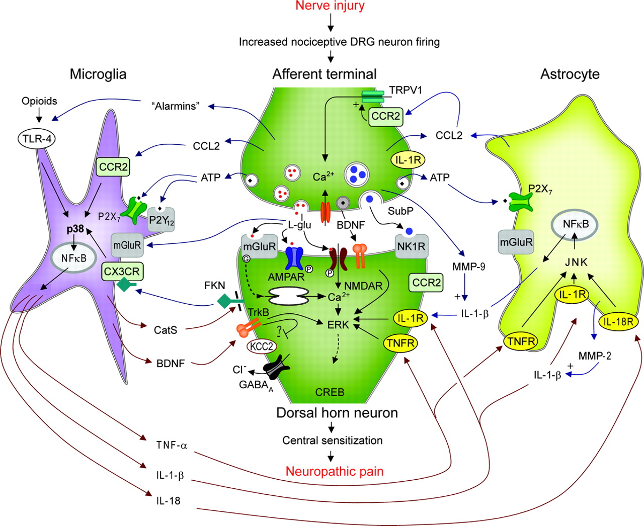
Signalling-MG-.jpg
 

Παρακαλώ συνδεθείτε ή εγγραφείτε για να απενεργοποιήσετε τις διαφημίσεις.

GOMI

Moderator
Μέλος του προσωπικού
weznem είπε:
κανε ενα περιληπτικο review στα ελληνικα οταν μπορεις γκομι.. :)

Είπα ότι σε έρευνα φάνηκε πως οι περιοχές του εγκεφάλου που έχουν τους περισσότερους υποδοχείς ινσουλίνης δείχνουν ότι είναι χωρισμένες σε 3 τομείς που ονομάζονται cortex. Οι περιοχές αυτές, όταν ο μεταβολισμός της γλυκόζης διαταραχθεί, τότε μέσω του υποθαλάμου υποκινούν τον οργανισμό να διασπάσει λιπαρά οξέα από τα λιποκύτταρα και να τα απελευθερώσει στο αίμα ώστε να πάρουν την απαιτούμενη ενέργεια από αυτά. Με αυτό το μηχανισμό έχουμε αυξημένη ροή λιπαρών οξέων στην περιοχή του εγκεφάλου.

Επειδή την εικόνα που είχα βάλει δεν την ανοίγει, δες εδώ πως φαίνονται αυτές οι περιοχές.

1379367760_sch%20drugs%20brain%20tissue.jpg


Η πορτοκαλί περιοχή (μετωπιαίος λοβός) είναι από τις πρώτες και ακολουθούν η μπλε (βρεγματικός λοβός) και η πράσινη (ινιακός λοβός).
Η κίτρινη περιοχή (κροταφικός λοβός) και αυτό που φαίνεται σαν καρύδι (cerebellum), δεν αναπτύσσουν ινσουλινοαντίσταση και η ανταπόκρισή τους στα λιπαρά οξέα είναι πολύ μειωμένη σε σχέση με τις άνω περιοχές. Το pattern της φαλάκρας, ακολουθεί ακριβώς το σχήμα των περιοχών αυτών (πορτοκαλί, μπλε, πράσινο) του εγκεφάλου. (επίσης εχθές παρατήρησα σε εντελώς φαλακρά κεφάλια, υπάρχει και η κλασσική μυτούλα που φαίνεται να κάνει η πίσω πράσινη περιοχή του εγκεφάλου)

Τώρα, υπάρχουν κάποια κύτταρα, που λέγονται Μικρογλοία (Microglia). Αυτά είναι εξειδικευμένα ανοσοκύτταρα του Κ.Ν.Σ, δηλαδή μακροφάγα που βρίσκονται σε διάφορες περιοχές του εγκεφάλου, κυρίως των άνω περιοχών. (grey matter) Η πορτοκαλί γραμμή που φαίνεται να ανεβαίνει προς τον εγκέφαλο, είναι ο νωτιαίος μυελός. (ΚΝΣ)

Είναι η πρώτη γραμμή άμυνας του εγκεφάλου και μιας και είναι άμεσα συνδεδεμένα με το κεντρικό νευρικό σύστημα, αντιδρούν άμεσα σε όποια ανωμαλία παρουσιαστεί στον οργανισμό υποκινώντας την αύξηση της προσταγλανδίνης D2 ώστε να προσελκύσει και άλλα μακροφάγα του οργανισμού μέσα από το αίμα προς το εγκέφαλο και ενεργοποιώντας τα Τ cells, δημιουργούν μια ασπίδα προστασίας στους ευαίσθητους νευρικούς ιστούς του εγκεφάλου.
Στην πίσω σελίδα φαίνεται πως και σε ποιες περιπτώσεις αντιδρούν.

Οπότε, μια αιτία θα μπορούσε να ήταν η διαταραχή της γλυκόζης, που αφορά και τοπικά στον εγκέφαλο αλλά και γενικά όλο τον οργανισμό άρα και την ενεργοποίηση της άμυνας του ΚΝΣ. H άλλη, αφορά δυστυχώς οτιδήποτε μπορεί να διαταράξει το ΚΝΣ. Λαβύρινθος δηλαδή.

Δεν ξέρω πόσο τραβηγμένο φαίνεται, αλλά μπορεί να εξηγήσει τα αυξημένα επίπεδα της pgd2 που βρέθηκε στα φαλακρά κεφάλια, μπορεί να εξηγήσει τα ενεργοποιημένα T cells που επίσης βρέθηκαν, μπορεί αν εξηγήσει το pattern που χάνουμε μαλλιά μόνο σε nw6 περιοχή και μπορεί να εξηγήσει και όλες τις άλλες αιτίες τριχόπτωσης που αφορούν απειλή-διαταραχή του ΚΝΣ, καθώς η αντίδρασή τους είναι έντονη (acute), όπως σε κάποιες περιπτώσεις τριχόπτωσης που αφορούν έντονο στρες-σοκ, ή χρόνια όπως η mpb, όπου ακόμα και σε αυτή, έχουμε δει πως οι αιτίες μπορεί να είναι διαφορετικές στον καθένα, αλλά η φαλάκρα ακολουθεί το ίδιο μοτίβο.

Δηλαδή, διαφορετική αιτία που απειλεί το ΚΝΣ (υπέρταση, διαβήτης, ινσουλινοαντίσταση, πολυκυστικές, εμμηνόπαυση, στρες, κατάθλιψη, ορμονική διαταραχή κ.α), αλλά το ίδιο πράγμα αντιμετωπίζουμε όλοι στο μαλλί.
Φαίνεται πως όσο περισσότερο μείνουν ενεργοποιημένα, τόσο περισσότερη ζημιά κάνουν σε γειτονικά κύτταρα, όπως πχ. τα προγονικά.


Επίσης, αν θέλουμε να δώσουμε και μια πρόχειρη εξήγηση των γονιδίων που συμμετέχουν σε αυτό, θα άρχιζα από εδώ

Androgen-mediated immune function is altered by the apolipoprotein E gene.
Brown CM1, Xu Q, Okhubo N, Vitek MP, Colton CA.
Author information

Abstract

Androgens, like estrogens, have been linked to neuroprotective effects in the brain and to the improvement of cognitive function.

Part of this effect may be due to the action of androgens on the innate immune response.

We have examined the action of dihydrotestosterone (DHT) and testosterone on immune activation in primary cultures of microglia, the central nervous system macrophage.

Our data indicate that DHT acts as an antiinflammatory agent and depresses both nitric oxide and TNFalpha production in a dose-dependent fashion.

However, testosterone treatment of microglia and peritoneal macrophages increased supernatant nitrite levels, indicative of a proinflammatory effect.

Because the apolipoprotein E (APOE) genotype also dramatically impacts macrophage function and has been linked to neurodegenerative disease, we compared the effects of APOE genotype on androgen-mediated regulation of inflammation using targeted replacement mice expressing only the human APOE3 or human APOE4 gene.

Our data show that the antiinflammatory activity of DHT is significantly reduced in APOE4 targeted replacement mice compared to APOE3 mice.

The effect was not due to an APOE isoform-specific change in androgen receptor mRNA and protein expression. Rather, innate immune signaling pathways regulated by androgens are altered in the APOE4 microglia.

Compared to APOE3 microglia, DHT treatment did not reduce the phosphorylation of p38 MAPK or p54/p56 Janus kinase in APOE4 mice.

Thus, our data suggest that DHT modulation of kinase activity is altered in microglia from mice expressing an APOE4 genotype and may impact androgen treatment therapies in individuals with an APOE4 genotype.

Απ ότι φαίνεται, σε κάποιους η dht δρα ως αντιφλεγμονώδη (apoe3 gene) μέσα από τα Μικρογλοία και σε κάποιους άλλους όχι (apoe4 gene).

Και λέω εγώ τώρα, αν δούμε την ινσουλίνη, όσο λιγότερο ανταποκρίνονται οι ιστοί μας σε αυτή, τόσο ο οργανισμός την αυξάνει ώστε να λάβουμε τα οφέλη της. Μήπως η dht που λένε ότι υπερεκφράζεται στα φαλακρά κεφάλια έχει έναν παρόμοιο μηχανισμό?
Δηλαδή φαίνεται ότι δρα προστατευτικά στα κύτταρα του ανοσοποιητικού ως αντιφλεγμονώδη, αλλά σε όσους φέρουν το γονίδιο apoe4, η δράση της είναι εξασθενημένη. Άρα μήπως ο οργανισμός την αυξάνει στην περιοχή, αλλά επειδή η δράση της λόγω του συγκεκριμένου γονιδίου έχει μεταβληθεί, κάνει απλά τα πράγματα χειρότερα....?
 

weznem

Well-Known Member
ευχαριστω γκομι για τον χρονο σου.. τελικα η φαλακρα μαλλον θα οδηγησει στο να μαθουμε-επιλύσουμε και αλλα προβληματα στον ανθρωπινο οργανισμο.. ισως τελικα αποδειχθεί οτι δεν ειναι ενα προβλημα αμιγώς αισθητικό..αλλα ενα προβλημα που κρουει τον κώδωνα του κινδύνου προτού εμφανιστουν πολλα προβληματα..
 

GOMI

Moderator
Μέλος του προσωπικού
The researchers examined the involvement of microglia brain cells in the development of depression following chronic exposure to stress. Comprising roughly 10% of brain cells, microglia are the representatives of the immune system in the brain; but recent studies have shown that these cells are also involved in physiological processes not directly related to infection and injury, including the response to stress.”

The researchers mimicked chronic unpredictable stress in humans - a leading cause of depression - by exposing mice to repeated, unpredictable stressful conditions over a period of 5 weeks.”

….”The researchers found that during the first week of stress exposure, microglia cells undergo a phase of proliferation and activation, reflected by increased size and production of specific inflammatory molecules, after which some microglia begin to die.

Following the 5 weeks of stress exposure, this phenomenon led to a reduction in the number of microglia, and to a degenerated appearance of some microglia cells, particularly in a specific region of the brain involved in responding to stress.

When the researchers blocked the initial stress-induced activation of microglia with drugs or genetic manipulation, they were able to stop the subsequent microglia cell death and decline, as well as the depressive symptoms and suppressed neurogenesis.

However, these treatments were not effective in "depressed" mice, which were already exposed to the 5-weeks stress period and therefore had lower number of microglia. Based on these findings, the investigators treated the "depressed" mice with drugs that stimulated the microglia and increased their number to a normal level.”

“…. "We were able to demonstrate that such microglia-stimulating drugs served as effective and fast-acting antidepressants, producing complete recovery of the depressive-like behavioral symptoms, as well as increasing the neurogenesis to normal levels within a few days of treatment.” “

This is the part that the supplements ubiquinol and PQQ may do from some to a lot in some cases. Ubiquinol helps your mitochondria stay healthy to begin with and PQQ has been found in studies at the University of California, Davis to cause new mitochondria to grow.


If this does indeed cause fewer microglial cells to die or stop functioning well or help BDNF from exercise and taking or eating DHA to build new and healthy microglial cells, it does look likely to stop this cause of depression.


Docosahexaenoic acid modulates inflammatory and antineurogenic functions of activated microglial cells.

Antonietta Ajmone-Cat M1, Lavinia Salvatori M, De Simone R, Mancini M, Biagioni S, Bernardo A, Cacci E, Minghetti L.
Author information

Abstract

The complex process of microglial activation encompasses several functional activation states associated either with neurotoxic/antineurogenic or with neurotrophic/proneurogenic properties, depending mainly on the extent of activation and the nature of the activating stimuli.

Several studies have demonstrated that acute exposure to the prototypical activating agent lipopolysaccharide (LPS) confers antineurogenic properties upon microglial cells.

Acutely activated microglia ortheir conditioned media (CM) reduce neural stem progenitor cell (NPC) survival and prevent NPC differentiation into neurons.

The present study tested the hypothesis that docosahexaenoic acid (DHA), a long-chain polyunsatured fatty acid (L-PUFA) with potent immunomodulatory properties, could dampen microglial proinflammatory functions and modulate their antineurogenic effect.

We demonstrate that DHA dose dependently inhibits the synthesis of inflammatory products in activated microglia without inducing an alternative antiinflammatory phenotype.

Among the possible DHA mechanisms of action, we propose the inhibition of p38 MAPK phosphorylation and the activation of the nuclear receptor peroxisome proliferator activated receptor (PPAR)-γ.

The attenuation of M1 proinflammatory phenotype has relevant consequences for the survival and differentiation of NPC, because DHA reverses the antineurogenic activities of conditioned media from LPS-activated microglia.

Our study identifies new relevant potentially protective and proneurogenic functions of DHA, exerted through the modulation of microglial functions, that could be exploited to sustain or promote neuroregenerative processes in damaged/aged brain. :dbann
 

GOMI

Moderator
Μέλος του προσωπικού
Hyperinsulinemia

The insulin sequence is:

Elevated insulin---increased delta desaturase enzymes---increased conversion of omega 6 fatty acids to arachidonic acid--increased prostaglandin 2's--increased production of cytokines--increased inflammatory response throughout the entire body.

The glucose sequence is:

Elevated serum glucose--increased intestinal candida--migration of candida to upper intestine--root formation of candida in duodenum--migration of candida spores throughout the bloodstream--candida infection throughout the body.

This candida source of infection is not the only source of infection. Because of the increased availability of cytokines, he body becomes subject to infection from many other sources. This is one reason that diabetics experience poor wound healing. One of the most important sites of infection is the coronary artery. This is located in the high pressure, sugar rich side of the blood supply. When the infection that occurs here is noticed by the immune system, the body attempts to patch the damage with cholesterol, Lp(a), free calcium and a few other things.

This patching process is known as atherosclerosis. As the patching process continues, the interior of the artery becomes narrowed. As the artery narrows, we become more susceptible to ischemic events. These are events where a clot forms in the artery and blocks the artery at a narrow atherosclerotic point. When this occurs in the coronary artery, the blood supply to the heart muscles is stopped and a heart attack ensues. This whole process is exacerbated by the fact that without adequate omega 3 fatty acids in the diet the blood thickens and the platelets become sticky and prone to form clots.

Notice the role of omega 6 fatty acids in making the entire body subject to the inflammatory response mediated by the prostaglandin 2 family of prostaglandins.

The body would, if it could, manufacture the antagonists to these prostaglandin 2's to prevent setting up this whole body inflammatory response. These antagonists are the prostaglandin 1's and 3's.

Our bodies, typically, cannot manufacture these anti-inflammatory prostaglandins because they lack the basic raw materials. These are the omega 3 fatty acids. We typically need and do not have, any omega 3 fatty acids in our diet at all. We have an over abundance of omega 6's and transfats in our diet. The separate damaging role of transfat substitution for omega 3 fatty acids has been discussed elsewhere in connection with the glucose transport system.

Νομίζω πως οι όλο και αυξανόμενοι ρυθμοί της τριχόπτωσης από μικρότερη ηλικία όπως επίσης και η αύξηση της γυναικείας τριχόπτωσης, έχει να κάνει σε πολύ μεγάλο βαθμό με την υπέρογκη καθημερινή κατανάλωση ω6 λιπαρών που απλά βρίσκονται παντού.

Για όποιον ενδιαφέρεται...

To stop and reverse the progress of Hyperinsulinemia the following dietary steps are mandatory:

Do not eat any hydrogenated oils or any prepared foods that contain them as ingredients.
Do not eat any unsaturated or refined vegetable oil that comes in a transparent bottle on a room temperature grocery store shelf. Cold pressed olive oil, coconut oil and Sesame seed oil are the only exceptions to this blanket rule.
Do not eat any oil that has been used in deep frying or that has been heated in cooking except butter and coconut oil. Do not eat any deep fried foods at all.
Add one or two tablespoons full of cold pressed flax oil or fish oil to the daily diet. Do not cook with this oil. Keep it refrigerated when not in use.
Supplement the diet with a good vitamin and chelated mineral complex from a reputable manufacturer. If you don't know one, get a recommendation from your local Naturopath or Chiropractor. Be sure to get Chromium, Vanadium, Calcium, Magnesium, and the trace minerals in the mix.
The program outlined above has a track record of reversing Hyperinsulinemia and the Type 2 diabetes that often accompanies it. In our Special Report, Insulin: Our Silent Killer, we are able to expand, explain and elaborate upon this protocol and present useful tricks and techniques to assure its speedy success in the vast majority of cases.

Select foods with a low Glycemic Index from a glycemic table. Our Special Report contains a Glycemic Index with instructions for use and much more information on this. Do not depend upon food exchanges; they don't work. They are based on the idea that food may be exchanged on the basis of equal calorie content and ignore that fact that, while calories are important, the glycemic value is far more important to the diabetic.
Use fiber in the diet as much as practical. It reduces the "rate effect" of Blood Sugar Control System perturbation and helps keep post prandial Glucose in check. Our Special Report elaborates upon the types of fiber and on how and why fiber works to control blood sugar.
Develop the habit of exercise at least twice a day. Work for twenty minutes or until you begain to perspire. Do not do aerobic exercises if your Arterial system has been compromised. Our Special Report discusses the additional non-Insulin dependent pathway that exercise opens for Glucose disposal.
Investigate the use of herbs such as Gymnema Sylvestre to help keep blood glucose low. We have more on this Herb as well as over thirty additional hypoglycemic herbs in our Special Report.
Test for allergic reactions when you eat by taking your pulse. When you eat a food to which you are allergic, your pulse rate will escalate at least 10 beats per minute. Don't eat any food to which you are allergic. This technology, too, is expanded and elaborated upon in our Special Report.
As a last resort, consult with your doctor regarding the use of Vanadium Sulfate and other oral hypoglycemic synthetic drugs. Be sure that you understand the side effects of these before you commit to using them. Our Special Report, Insulin: Our Silent Killer discusses rather thoroughly the pros and cons of these medications. It is very useful information to have if you need to discuss these issues with your physician.
In the beginning of the recovery program is is best to remove fats and oils, except flax seed oil, or other oil containing a high percentage of W3 fatty acids, from the diet as completely as possible. Later, when the Hyperinsulinemia has started to reverse, it is ok to gradually restore fats and oils, good fats and oils only, to as much as fifteen percent of the calories consumed.

This program will, relatively quickly, reverse Hyperinsulinemia, Type 2 diabetes and some of the other symptomatic diseases caused by Hyperinsulinemia. In my case, my Type 2 diabetes was reversed in 103 days from start to finish. At the start my fasting blood sugar was 368mg/dl. At the end of the program it varied between 75 and 85 mg/dl.

This program will remove much stress from the components of the Blood Sugar Control System; over a period of time they too will be restored to youthful function. This includes the Liver, Pancreas, Adrenals, Thyroid and the interior transport agents in each cell of our body.

This program will slow and in some cases reverse Vascular damage and Gangrenous damage to our extremities. There are faster ways to reverse this sort of damage which is thoroughly discussed in our Special Report.

This program will, over time, revcrse much of the Neuropathic damage to the nervous system.

It will reverse Atherosclerosis too, but may take years to do it. But here too, there are faster and better ways to reverse Atherosclerosis which we cover in our Special Report.
 

giannis25

Member
άρα εκτος του οτι το epa εμποδίζει την pgd2 και το dha εμποδίζει τις φλεγονές. γι αυτο παρατηρούμε όλοι μειωμένη τριχόπτωση μόνο 3,4 τρίχες.
 

GOMI

Moderator
Μέλος του προσωπικού
Πάνω κάτω φαίνεται πως συνεργάζονται η αυξημένη ινσουλίνη με τα ω-6 λιπάρα στο θέμα φλεγμονής. Τα ω-3, ενώ δεν επεμβαίνουν άμεσα στο θέμα ινσουλίνη-γλυκόζη, υποκινούν την παραγωγή "καλών" προσταγλανδινών που έχουν αντιφλεγμονώδη δράση όπως οι pge1 k pge3. Δείχνουν δηλαδή ότι σε μεγάλο βαθμό μαζεύουν το χάλι που προκαλεί η ινσουλινοαντίσταση.

Μοιάζει ανθρωπίνως αδύνατον πάντως να καταφέρει κάποιος, ειδικά στον κόσμο που ζούμε, να κρατήσει το ratio 2:1 (ω6/ω3)
 

GOMI

Moderator
Μέλος του προσωπικού
kapa, (ή όποιος άλλος φίλος ενδιαφέρεται) επειδή ξέρεις από ποια οπτική γωνιά πρέπει να ψάχνεις ένα συμπλήρωμα που να βοηθάει στην Α.Α. ρίχνε και μια ματιά σε ότι αφορά τα microglial cells.
Επειδή ο χρόνος της θεραπείας που κάνω για τις ορμόνες και την ινσουλίνη με πιέζει, ρίχνω όλο τον ελεύθερο χρόνο μου εκεί, αναζητώντας τρόπους να αποφύγω την εξωγενή χορήγηση ορμονών. Ο γιατρός μου έδωσε περιθώριο 2 μηνών και έχω ήδη διανύσει πάνω από 2 εβδομάδες.

Δεν μπορώ να είμαι 100% σίγουρος ακόμα για αυτά τα κύτταρα που βρίσκονται σε κάποιες περιοχές του εγκεφάλου, αλλά δες έναν μηχανισμό της ασταξανθίνης στη δράση τους.

Effects of astaxanthin on the production of NO and the expression of COX-2 and iNOS in LPS-stimulated BV2 microglial cells.

Choi SK1, Park YS, Choi DK, Chang HI.

Author information

Abstract

Astaxanthin has shown antioxidant, antitumor, and antiinflammatory activities; however, its molecular action and mechanism in the nervous system have yet to be elucidated.

We examined the in vitro effects of astaxanthin on the production of nitric oxide (NO), as well as the expression of inducible NO synthase (iNOS) and cyclooxygenase-2 (COX-2) in lipopolysaccharide (LPS)-stimulated BV2 microglial cells.

Astaxanthin inhibited the expression or formation of nitric oxide (NO), iNOS and COX-2 in lipopolysaccharide (LPS)-stimulated BV-2 microglial cells.

Astaxanthin also suppressed the protein levels of iNOS and COX-2 in LPS-stimulated BV2 microglial cells.

These results suggest that astaxanthin, probably due to its antioxidant activity, inhibits the production of inflammatory mediators by blocking iNOS and COX-2 activation or by the suppression of iNOS and COX-2 degradation.

Όλα είναι εκεί, pgd2, cox-2, inos, NO, ελεύθερες ρίζες, tnf-a, IL-1-6.

Οι λιποπολυσακχαρίτες (LPS) είναι μεγάλα μόρια, αποτελούμενα από ένα λιπίδιο κι έναν πολυσακχαρίτη ενωμένα με ομοιοπολικό δεσμό. Συνήθως βρίσκονται στην εξωτερική μεμβράνη του κυτταρικού τοιχώματος των gram-αρνητικών βακτηρίων, συμπεριλαμβανομένων των κυανοβακτηρίων, όπου δημιουργούν συμπλέγματα με πρωτείνες και φωσφολιπίδια. Οι λιποπολυσακχαρίτες συμπεριφέρονται ως ενδοτοξίνες και μπορούν να προκαλέσουν ερεθισμό και αλλεργικές αντιδράσεις όταν έρθουν σε επαφή με κάποιο ιστό. Σε σχέση με τους λιποπολυσακχαρίτες άλλων παθογόνων gram-αρνητικών βακτηρίων, όπως η Salmonella, οι λιποπολυσακχαρίτες των κυανοβακτηρίων δεν έχουν τόσο ισχυρή τοξική δράση.

http://jem.rupress.org/content/185/9/1661

Επίσης επειδή όπως σου είπα υπάρχει μεγάλη περίπτωση σε 2 μήνες να χρειαστεί να παίξω εξωγενή τέστο και επειδή όπου κοιτάξω, σε έχοντες και μη έχοντες πρόβλημα φαλάκρας, η τεστοστερόνη τους προκάλεσε τριχόπτωση, είδα πως δρα αυξάνοντας τα νιτρικά σε αυτά κύτταρα προκαλώντας την φλεγμονώδη αντίδρασή τους.
Ήδη ένα μέλος στο immortal που ξεκίνησε TRT, το πρώτο πράγμα που ένιωσε ήταν φλεγμονή στο κεφάλι, αν και δεν τον ένοιαζε ιδιαίτερα γιατί ήταν ήδη φαλακρός, αλλά είπε πως αν είχε μαλλιά σίγουρα θα τα μάζευε από το πάτωμα.

Επίσης ένα φλαβονοειδή που λέγεται baicalein, έχει μια πολύ σημαντική δράση απέναντι σε αυτά τα κύτταρα.

Inhibition of microglial activation by the herbal flavonoid baicalein attenuates inflammation-mediated degeneration of dopaminergic neurons

Morphological study shows that baicalein (5 µM) almost completely blocked LPS-induced activation of microglia. Excessive production of TNFα and free radicals such as NO and superoxide by LPS stimulation were also attenuated by baicalein at a concentration-dependent pattern.

Περισσότερα εδώ
http://link.springer.com/article/10.1007...004-0213-0

Επίσης τοπικό με αυτό το συστατικό κυκλοφορεί ήδη για την τριχόπτωση.

http://www.centre-clauderer.com/en/clauderer-treatments/baicalein.htm

Η φλεγμονώδη αντίδραση αυτών των κυττάρων δημιουργεί συνθήκες φαγοκυττάρωσης γι αυτό όσο περισσότερο μένουν ενεργοποιημένα, τόσο δυσκολότερο, εως αδύνατο γίνεται η υπόθεση regrowth μιας και βασικά κύτταρα όπως τα progenitor μαζί με τα dermal papillae καταστρέφονται.


Αν η δουλειά μου ήταν στο χώρο της έρευνας, θα ξεσκόνιζα πολύ προσεκτικά αυτόν τον μηχανισμό γιατί ποτέ και καμία έρευνα δεν στράφηκε σε αυτά.
Δυστυχώς έχω πολλά προβλήματα αυτόν τον καιρό για να μπορέσω να ασχοληθώ ιδιαίτερα, αλλά νομίζω τοπική astaxanthin (θέλει λίγο ψάξιμο τι γίνεται με τον ήλιο) μαζί με baicalein + tocotrienols θα ήταν πολύ καλό τοπικό για ΠΡΟΛΗΨΗ και όραλ μέχρι στιγμής φαίνεται ότι βοηθούν DHA, Ubiquinol, PQQ.
Για regrowth θέλουμε κάτι που να δημιουργεί ή έστω να αυξήσει τα dp cells.
 

GOMI

Moderator
Μέλος του προσωπικού
kapa είπε:
GOMI είπε:
kapa, (ή όποιος άλλος φίλος ενδιαφέρεται) επειδή ξέρεις από ποια οπτική γωνιά πρέπει να ψάχνεις ένα συμπλήρωμα που να βοηθάει στην Α.Α. ρίχνε και μια ματιά σε ότι αφορά τα microglial cells.
μπορω να απαντησω με σιγουρια μονο σε αυτο.
ο συνδιασμος 500mg DHA+500mg curcumin+200mg resveratrol θα ξεπερασει τα αποτελεσματα της φιναστεριδης.
η φαλακρα ειναι τοσο πολυδιαστατη που οσο την ψαχνω χανομαι.
για τις 3 ουσιες που προανεφερα ειμαι σιγουρος.
μια,μια θα τις προσθεσω

Είναι πολυδιάστατη κατάσταση ως προς το τι μπορεί να προκαλέσει την αντίδραση των ανοσοκυττάρων του Κ.Ν.Σ.
Αυτό μπορεί να το κάνουν πάρα πολλά πράγματα από τα οποία κάποια γονίδια μας δεν μας προστατεύουν εξ αρχής.
Μετά από αυτό όμως, η δράση των microglial cells, είναι ξεκάθαρη και ίδια σε όλους. Εξάλλου ουσιαστικά πρόκειται για μακροφάγα κύτταρα.
Και η κουρκουμίνη και η ρεσβερατρόλη πάνω σε αυτά δουλεύουν.

Curcumin is a potent modulator of microglial gene expression and migration

Microglial cells are important effectors of the neuronal innate immune system with a major role in chronic neurodegenerative diseases. Curcumin, a major component of tumeric, alleviates pro-inflammatory activities of these cells by inhibiting nuclear factor kappa B (NFkB) signaling.

To study the immuno-modulatory effects of curcumin on a transcriptomic level, DNA-microarray analyses were performed with resting and LPS-challenged microglial cells after short-term treatment with curcumin.

Curcumin treatment markedly changed the microglial transcriptome with 49 differentially expressed transcripts in a combined analysis of resting and activated microglial cells.

Curcumin effectively triggered anti-inflammatory signals as shown by induced expression of Interleukin 4 and Peroxisome proliferator activated receptor α. Several novel curcumin-induced genes including Netrin G1, Delta-like 1, Platelet endothelial cell adhesion molecule 1, and Plasma cell endoplasmic reticulum protein 1, have been previously associated with adhesion and cell migration. Consequently, curcumin treatment significantly inhibited basal and activation-induced migration of BV-2 microglia. Curcumin also potently blocked gene expression related to pro-inflammatory activation of resting cells including Toll-like receptor 2 and Prostaglandin-endoperoxide synthase 2. Moreover, transcription of NO synthase 2 and Signal transducer and activator of transcription 1 was reduced in LPS-triggered microglia. These transcriptional changes in curcumin-treated LPS-primed microglia also lead to decreased neurotoxicity with reduced apoptosis of 661W photoreceptor cultures.

Conclusions
Collectively, our results suggest that curcumin is a potent modulator of the microglial transcriptome.
Curcumin attenuates microglial migration and triggers a phenotype with anti-inflammatory and neuroprotective properties. Thus, curcumin could be a nutraceutical compound to develop immuno-modulatory and neuroprotective therapies for the treatment of various neurodegenerative disorders.

http://www.jneuroinflammation.com/content/8/1/125


Resveratrol inhibits nitric oxide and TNF-alpha production by lipopolysaccharide-activated microglia.
Bi XL1, Yang JY, Dong YX, Wang JM, Cui YH, Ikeshima T, Zhao YQ, Wu CF.
Author information
Abstract
Upon activation, brain macrophages, the microglia, release proinflammatory mediators that play important roles in eliciting neuroinflammatory responses associated with neurodegenerative diseases.

As resveratrol, an antioxidant component of grape, has been reported to exert anti-inflammatory activities on macrophages, we investigated its effects on the production of TNF-alpha (TNF-alpha) and nitric oxide (NO) by lipopolysaccharide (LPS)-activated microglia.

Exposure of cultured rat cortical microglia and a mouse microglial cell line N9 to LPS increased their release of TNF-alpha and NO, which was significantly inhibited by resveratrol. Further studies revealed that resveratrol suppressed LPS-induced degradation of IkappaBalpha, expression of iNOS and phosphorylation of p38 mitogen-activated protein kinases (MAPKs) in N9 microglial cells. These results demonstrate a potent suppressive effect of resveratrol on proinflammatory responses of microglia, suggesting a therapeutic potential for this compound in neurodegenerative diseases accompanied by microglial activation.
 
Μπλουζα